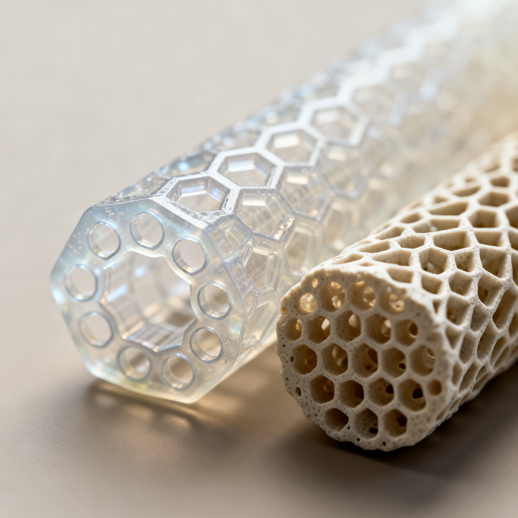
Custom scaffold: the geometry that regenerates?
3D printing does not merely copy the shape of tissues: thanks to intelligent geometries and materials studied in minute detail, it is reinventing the actual supports on which cells grow. Two recent patents show how hexagonal microchannels and gyroid structures are transforming nerve and bone scaffolds into more effective devices, without abandoning already known and approved materials.
- BIOMIMETIC SCAFFOLD FOR PERIPHERAL NERVE INJURIES — January 14, 2026
- METHOD FOR PRODUCING A 3D PRINTED BONE GRAFT AND 3D PRINTED BONE GRAFT — 13 May 2026
Geometries that guide growth
The new microstructural designs, such as hexagonal and gyroid, improve cell alignment and distribution of biochemical signals. They are not random shapes, but architectures designed to influence cell behavior.
The patent for peripheral nerve scaffolds introduces a hexagonal microchannel structure that runs through the entire device. Each channel has an internal diameter between 150 µm and larger sizes, with thin walls that leave more open space compared to traditional circular channels.
The hexagonal geometry is not an aesthetic choice. According to the patent “Biomimetic Scaffold for Peripheral Nerve Injuries”, the hexagonal microchannels promote a more uniform alignment of regenerating axons. The hexagons are placed side by side without leaving gaps, creating a continuous network that guides nerve growth from the proximal to the distal stump.
The scaffold hosts from 7 to 200 microchannels, depending on the external diameter of the device. The length ranges from 0.5 to 15 cm, with an external diameter between 1.5 and 10 mm. At the ends, two “overhangs” allow suturing to the patient’s nerve tissue.
- Hexagonal microchannels with 150 µm inner diameter
- From 7 to 200 channels per device
- Length from 0.5 to 15 cm, outer diameter from 1.5 to 10 mm
- Overhangs at the ends for suturing to nerve tissue
For bone, the patent “Method for Producing a 3D Printed Bone Graft” proposes a gyroid structure. It is a triply periodic minimal surface (TPMS) that maximizes the surface-to-volume ratio. The gyroid achieves a ratio of 4.1, compared to 2.3 for Schwarz and 2.8 for Diamond.
This high ratio means more surface area available for cell adhesion and vascularization. Scaffolds printed with this geometry show an open porosity between 46% and 52%, with interconnected pores of about 600 µm. The distribution is measurable via micro-computed tomography (µ-CT), confirming the repeatability of the process.
Known materials, new results
The use of already approved materials such as PEGDA and GelMA allows a shorter path to market, while maintaining high mechanical and biological performance. The novelty lies not in the basic chemistry, but in the combination and the process.
The patent for neural scaffolds uses poly(ethylene glycol) diacrylate (PEGDA) and methacrylated collagen. The weight ratio ranges from about 125:1 to 25:1, with specific formulations at 100:1, 75:1, 50:1. These materials are biodegradable and already known in the medical field.
The structure can incorporate neurotrophic factors such as BDNF (brain-derived neurotrophic factor) or NGF (nerve growth factor). Controlled release occurs from the scaffold walls, stimulating axon growth during regeneration. In some variants, the channels can be filled with Schwann cells to further support the process.
For bone scaffolds, the patent describes ceramic formulations loaded in photopolymerizable resins. The compositions include PEGDA or GelMA as a polymer matrix, with calcium phosphate-based ceramic fillers. The formulations are identified as CAP40, CAP60, CAP80, CAP100 (with PEGDA) and CAG10 (with GelMA).
| Formulation | Base exposure (s) | Layer exposure (s) |
|---|---|---|
| CAP40 | 150 | 100 |
| CAP60 | 100 | 40 |
| CAP80 | 100 | 40 |
| CAP100 | 100 | 30 |
| CAG10 | 40 | 8 |
After printing, the bone scaffolds are cleaned with ultrasound in distilled water. PEGDA and GelMA are water-soluble, so the process removes unpolymerized monomer from the porous structure. The scaffolds are then immersed in water at 37°C to allow the transformation of ’α-TCP (alpha-tricalcium phosphate) into calcium-deficient hydroxyapatite (CDHA).
From theory to production
Companies can integrate these technologies using their existing production lines, but they face costs and times that are not yet optimized. Scalability depends on well-defined and repeatable process parameters.
The patent for nerve scaffolds describes an additive manufacturing process that can be implemented with already available 3D printing technologies. The geometry is designed from digital models, with precise control over the number, size, and arrangement of microchannels.
A company that produces nerve reconstruction devices could adopt this technology using its own 3D printing lines. The materials (PEGDA, methacrylated collagen) are commercially available and the process does not require completely new equipment. However, validation of printing parameters and post-processing protocols takes time.
For bone scaffolds, the patent “Method for Producing a 3D Printed Bone Graft” provides specific exposure parameters for each formulation. The layer thickness is set at 50 µm. Exposure times vary according to composition: CAP40 requires 100 seconds per layer, CAG10 only 8 seconds.
Bone scaffold production process
- Preparation of .stl file: import of the gyroid geometry into the slicing software.
- SLA printing: layer-by-layer production with optimized exposure parameters.
- Ultrasonic cleaning: removal of unpolymerized monomer in distilled water.
- Hardening: immersion in water at 37°C for transformation of α-TCP into CDHA.
An orthopedic device manufacturer could integrate this technology using SLA printers with already validated exposure parameters. Repeatability is confirmed by µ-CT measurements, which show porosity consistent with the design. However, production and cleaning times could limit its use in surgical emergency contexts.
Real limits and challenges
Long-term biocompatibility and regulatory validation processes still represent a bottleneck for large-scale adoption. Not all positive in vitro signals translate into clinical success.
The patent for nerve scaffolds does not provide clinical data on patients. The long-term biological response to hexagonal microchannels is still under study. It is not clear whether the hexagonal geometry maintains the advantages observed in vitro also under in vivo conditions, where factors such as inflammation, vascularization, and mechanical load can influence the outcome.
Production costs could be higher than current systems. 3D printing of scaffolds with complex geometries requires time and specific materials. Suturing the overhangs to the patient's nerve tissue requires precise surgical skills.
The patent “Biomimetic Scaffold for Peripheral Nerve Injuries” does not specify material degradation times nor provide direct comparisons with existing commercial devices.
For bone scaffolds, the patent “Method for Producing a 3D Printed Bone Graft” does not address the long-term biocompatibility of specific formulations. µ-CT analyses confirm the porous structure but do not provide data on cell adhesion, vascularization, or bone integration in animal models or patients.
The exposure parameters are specific to the printing device used. Variations in the machine or formulations require new optimizations. This limits the immediate transferability of the process to other production lines.
Pilot clinical studies on 3D-printed scaffolds for bone regeneration show good integration and volumetric increase of bone in dentistry and maxillofacial surgery. However, the clinical introduction of scaffolds with complex geometries still requires extensive preclinical and clinical testing, with attention to safety, degradation kinetics, and long-term mechanical reliability.
Smart geometries, patient adoption
New geometries and biocompatible materials are opening up concrete scenarios for more effective medical scaffolds. Hexagonal microchannels and gyroid structures are not just design exercises but attempts to influence cell behavior in a measurable way.
The use of already known materials such as PEGDA, GelMA, and methacrylated collagen reduces part of the regulatory risk. 3D printing processes are repeatable and parameters are documented. However, the transition from research to clinic requires extensive validations, investments in quality control, and not short times.
Companies that today begin to integrate these technologies into their processes will be those that tomorrow will lead the market for personalized implants. But adoption requires patience, multidisciplinary skills, and a clear understanding of current limitations.
article written with the help of artificial intelligence systems
Q&A
What is the advantage of hexagonal microchannels over circular ones in nerve scaffolds?
Hexagonal microchannels have thinner walls that leave more open space compared to circular ones. Additionally, hexagons fit side by side without empty spaces, creating a continuous network that promotes more uniform alignment of regenerating axons from the proximal stump to the distal stump.
What is a gyroid structure and why is it suitable for bone scaffolds?
The gyroid is a triply periodic minimal surface (TPMS) that maximizes the surface-to-volume ratio, reaching a value of 4.1. This high ratio provides more surface area for cell adhesion and vascularization, with an open porosity between 46% and 52% and interconnected pores of approximately 600 µm.
Which materials are used for these scaffolds and what is their main advantage?
Already approved and well-known medical-grade materials are used, such as PEGDA, GelMA, and methacrylated collagen. The use of these substances reduces regulatory risks and shortens the path to market, while maintaining high mechanical and biological performance.
What are the main challenges and limitations for the clinical adoption of these technologies?
Long-term biocompatibility and regulatory validation processes still represent a bottleneck. Patents do not provide clinical data on patients, and it is unclear whether the advantages observed in vitro are maintained in vivo, where inflammation and mechanical loading can influence the results.
What are the specific production parameters for the bone scaffolds described in the article?
The layer thickness is set to 50 µm, while exposure times vary depending on the formulation: for example, CAP40 requires 100 seconds per layer and CAG10 only 8 seconds. After printing, the scaffolds are cleaned with ultrasound in distilled water and immersed in water at 37°C to transform α-TCP into calcium-deficient hydroxyapatite (CDHA).