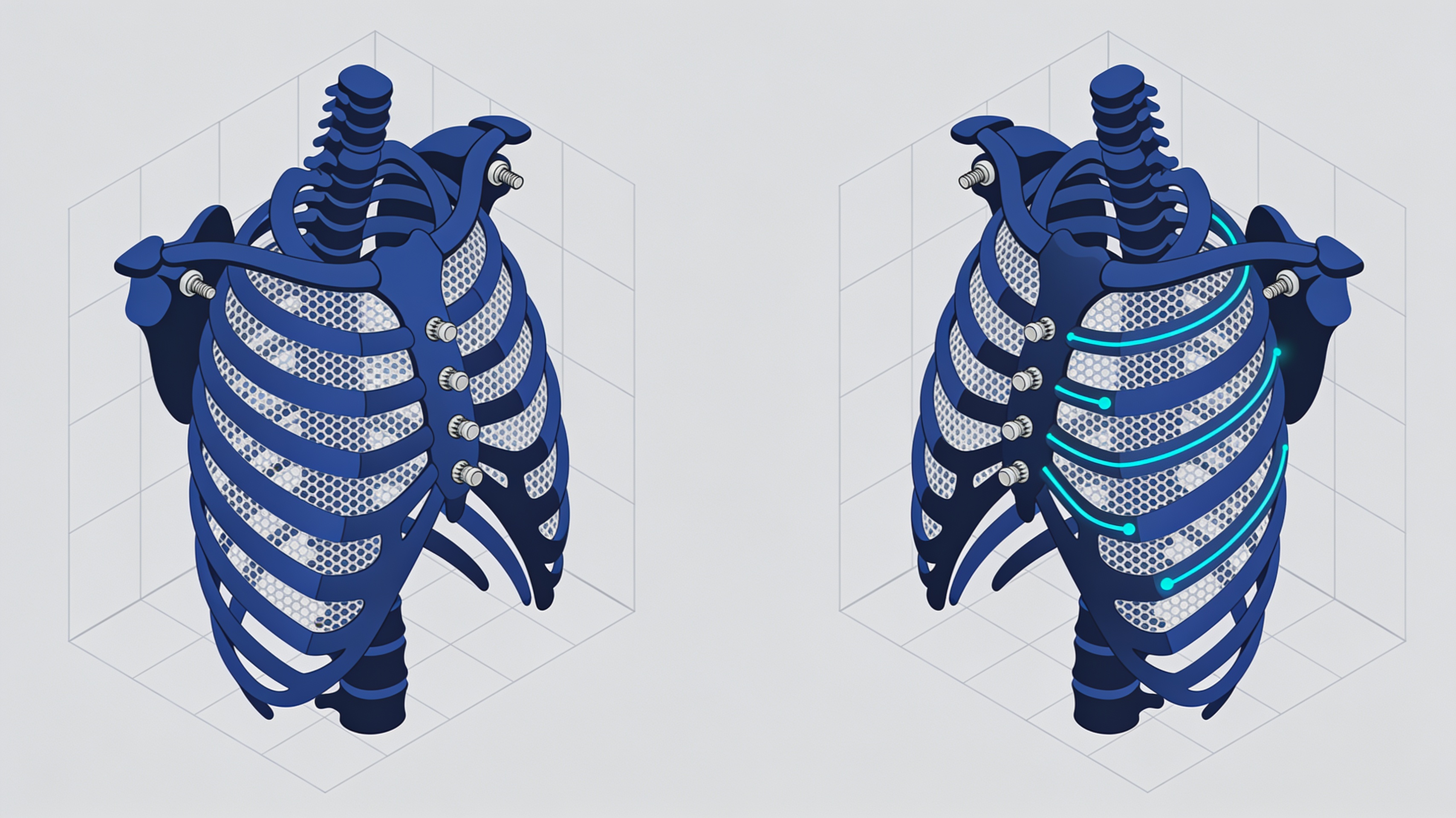
3D-printed titanium thoracic implants: the complete workflow from Morriston Hospital
Morriston Hospital in Swansea has formalized a complete digital chain to reconstruct large portions of the chest wall with custom titanium implants, produced using Laser Powder Bed Fusion. The study, published in September 2026, documents six consecutive patients who underwent extensive thoracic resections for malignant or radiotherapy-related conditions.
The most relevant finding is not only the use of metal 3D printing, already known in the literature. It is the standardization of a process that starts from CT and reaches the definitive implant without intraoperative modifications.
- Six patients treated without intraoperative modifications to the implants
- Follow-up from 5 months to 5 years without hardware failure
- Complete digital workflow: CT → segmentation → CAD → physical verification → printing
- Material: Ti-6Al-4V ELI Grade 23
The digital chain: from diagnostics to the implant
The workflow integrates medical imaging, virtual oncological planning, CAD design, and physical verification before final production.
The process begins with the patient's CT scan. The data are segmented to reconstruct the three-dimensional anatomy and virtually plan the oncological resection margins.
This is followed by CAD design of the implant, modeled on the specific anatomy. Before titanium production, the team creates 1:1 scale stereolithographic prototypes to physically verify geometry and fit.
Only after this physical validation does final printing proceed in Ti-6Al-4V ELI Grade 23 using Laser Powder Bed Fusion. This intermediate step reduces the risk of costly errors and ensures that the implant corresponds exactly to the surgical plan.
Why thoracic resections are so complex
Large resections simultaneously involve bone, cartilage, and soft tissue structures, making standardized reconstruction impossible.
A thoracic tumor can extend through ribs, costal cartilages, sternum, and muscles. The surgical priority remains oncologically adequate removal, but extensive resections create defects that compromise organ protection and respiratory stability.
In the six cases studied, all bone defects exceeded 5 cm and were not suitable for primary closure. Two patients required reconstruction of ribs only, two had composite defects involving ribs, costal cartilages, and partial sternum, and another two had extensive sternal resections.
The geometry to be reconstructed was therefore different in each case. This makes customization not an option, but a clinical necessity.
Operational workflow
- CT scan acquisition: Diagnostic imaging of the patient.
- Anatomical segmentation: 3D reconstruction of thoracic structures.
- Virtual planning: Definition of oncological resection margins.
- CAD design: Modeling of the custom implant.
- SLA prototype: Physical verification at 1:1 scale.
- L-PBF printing: Final production in titanium Ti-6Al-4V ELI Grade 23.
Clinical results and study limitations
No hardware failure during follow-up up to five years, but the limited case series requires validation on a larger scale.
During follow-up, ranging from five months to five years, no implant failures or complications directly related to the hardware were observed. In none of the six cases was intraoperative modification of the implant or resection plans required.
The finding is encouraging but must be contextualized. The authors themselves present the work as a proof-of-concept intended for validation through larger, multicenter studies.
Case series of six patients at a single center. Multicenter studies are needed to validate safety and efficacy in larger populations and with standardized follow-up.
Implications for medical additive manufacturing
The case demonstrates how mass customization requires standardization of the process, not of the product.
This workflow represents an example of mass customization applied to medical devices. Each implant is unique, but the process is standardized and repeatable.
Laser Powder Bed Fusion makes it possible to produce complex geometries that are impossible with traditional techniques. Ti-6Al-4V ELI Grade 23 guarantees biocompatibility and mechanical properties suitable for thoracic load-bearing.
Physical verification using stereolithographic prototypes before final production is a distinctive element. It reduces clinical risks and costs, allowing corrections while the material is still polymeric and not titanium.
The next step will be to demonstrate that this approach also works outside a single specialized center, with different surgeons and more heterogeneous case series.
article written with the help of artificial intelligence systems
Q&A
Which 3D printing technology does Morriston Hospital use for thoracic implants?
Morriston Hospital uses Laser Powder Bed Fusion to produce the final titanium implants. This is a laser melting technology on a metal powder bed, suitable for complex and customized geometries.
Which material is used for customized thoracic implants?
The implants are made of Ti-6Al-4V ELI Grade 23, a biocompatible titanium alloy with high mechanical properties. This material is widely used in the medical field for its strength and compatibility with tissues.
How many phases does the digital workflow described in the study involve?
The workflow starts from the patient's CT scan, then proceeds with segmentation, virtual oncological planning, and CAD design. Before the final titanium printing, the team produces 1:1 scale stereolithographic prototypes to verify geometry and fit.
Why was customization of the implant necessary in the studied cases?
All bone defects exceeded 5 cm and were not suitable for primary closure. The geometry to be reconstructed varied in each case, involving ribs, costal cartilages, and sternum, making a standardized solution impossible.
What clinical results were observed in the six treated patients?
The six patients were operated on without intraoperative modifications to the implants. Follow-up, ranging from 5 months to 5 years, showed no hardware failure.