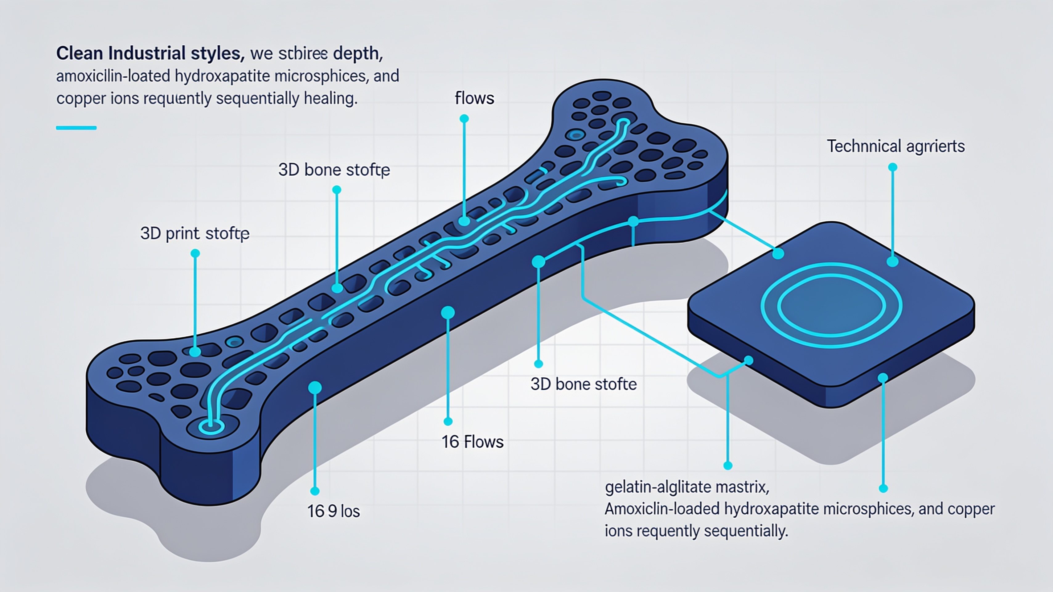
3D-printed bone scaffold that changes function during healing: the NTU Singapore research
Researchers at Nanyang Technological University have developed a scaffold for bone regeneration that releases antibiotic in the early stages and stimulates vascularization and bone growth at a later stage. The system combines gelatin, alginate, copper ions, amoxicillin, and hydroxyapatite in a single 3D-printed structure.
Large bone defects represent a complex clinical challenge. Unlike simple fractures, when trauma, infection, or tumor resection cause extensive loss of substance, the tissue cannot regenerate on its own.
The problem is not only mechanical. First, the bacterial infection must be controlled, then a vascular network for oxygen and nutrients must be developed, and finally a stable environment for bone regeneration must be created.
A sequential approach to healing
The research introduces the concept of “fast-then-slow” release, in which the same substances produce different biological effects at different times in the healing process.
An international group led by NTU Singapore has published a study in Materials Today Bio that presents a 3D-printed scaffold designed around this biological sequence. The work is authored by Yongteng Song, Haiguang Zhang, Yuxuan Tang, Jie Wang, Shujun Zhang, Xinchao Gao, Yuanhang Yang, Qingxi Hu, and Changjin Huang.
The structure combines a matrix of gelatin and sodium alginate with copper ions and porous hydroxyapatite microspheres loaded with amoxicillin. Each component has a specific role in the healing sequence.
- Matrix: gelatin and sodium alginate for the base structure
- Antibiotic: amoxicillin for controlling initial infections
- Vascularization: copper ions to stimulate blood vessel formation
- Regeneration: hydroxyapatite for bone growth
Beyond simultaneous release
The main innovation does not lie in the individual materials, but in their temporal orchestration, designed to follow the natural phases of bone healing.
Traditional scaffolds release all agents simultaneously, with no relation to the course of healing. The new approach instead aims for a “fast-then-slow” dynamic, in which the substances act in sequence.
The project involves NTU Singapore, Shanghai University, and other Chinese institutes. It is not yet a clinical implant, but a preclinical proof-of-concept tested in the laboratory and in an animal model of cranial defect in rats.
The clinical context of bone defects
Autografting remains the gold standard, but it has significant limitations that push research toward multifunctional synthetic alternatives.
Bone autografting remains one of the clinical references because the material harvested from the patient possesses structure, cells, and biological signals together. However, it requires a second surgical site, the available quantity is limited, and harvesting can cause pain and complications.
Allografts and synthetic materials avoid some problems, but they do not automatically recreate the entire environment necessary for regeneration. Hence the need for scaffolds that integrate multiple functions in sequence.
The work is published under the title “3D-printed multifunctional composite scaffolds incorporating copper ions/amoxicillin/hydroxyapatite for synergistic antibacterial and vascularized bone regeneration” in Materials Today Bio. This is preclinical research, not a device ready for human use.
Prospects for biomedical additive manufacturing
3D printing makes it possible to combine materials with different properties into complex geometries, opening new possibilities for bone tissue engineering.
The main interest of the project is conceptual. It demonstrates how additive manufacturing can integrate components with different release kinetics and biological functions into a single structure.
The ability to print scaffolds that respond to the temporal phases of healing represents a step forward compared with static systems. Efficacy remains to be verified in larger preclinical models and, possibly, in clinical studies.
The project illustrates how 3D printing in the biomedical field is not only replication of anatomical shapes, but the design of dynamic functional systems that interact with biological processes over time.
article written with the help of artificial intelligence systems
Q&A
Who developed the sequential 3D scaffold for bone regeneration?
The project is led by NTU Singapore with the collaboration of Shanghai University and other Chinese institutes. The work is signed by Yongteng Song, Haiguang Zhang, Yuxuan Tang, Jie Wang, Shujun Zhang, Xinchao Gao, Yuanhang Yang, Qingxi Hu, and Changjin Huang and is published in Materials Today Bio.
How does the fast-then-slow release of the scaffold work?
The scaffold first releases the antibiotic amoxicillin to control bacterial infection in the early stages. Subsequently, copper ions act to stimulate vascularization and hydroxyapatite to promote bone growth. This approach follows the natural biological sequence of healing, unlike traditional scaffolds that release all agents simultaneously.
What materials make up the 3D-printed scaffold?
The structure combines a gelatin and sodium alginate matrix with copper ions and porous hydroxyapatite microspheres loaded with amoxicillin. Each component has a specific role: the matrix provides the base structure, amoxicillin controls infections, copper stimulates blood vessels, and hydroxyapatite promotes bone regeneration.
Is the scaffold already available for clinical use?
No, it is a preclinical proof-of-concept tested in the laboratory and on an animal model of cranial defect in rats. It is not yet a clinical implant approved for human use.
Why do large bone defects require a multifunctional scaffold?
Unlike simple fractures, large bone defects from trauma, infection, or tumor resection cannot regenerate on their own. It is necessary first to control bacterial infection, then to develop a vascular network for oxygen and nutrients, and finally to create a stable environment for bone regeneration.