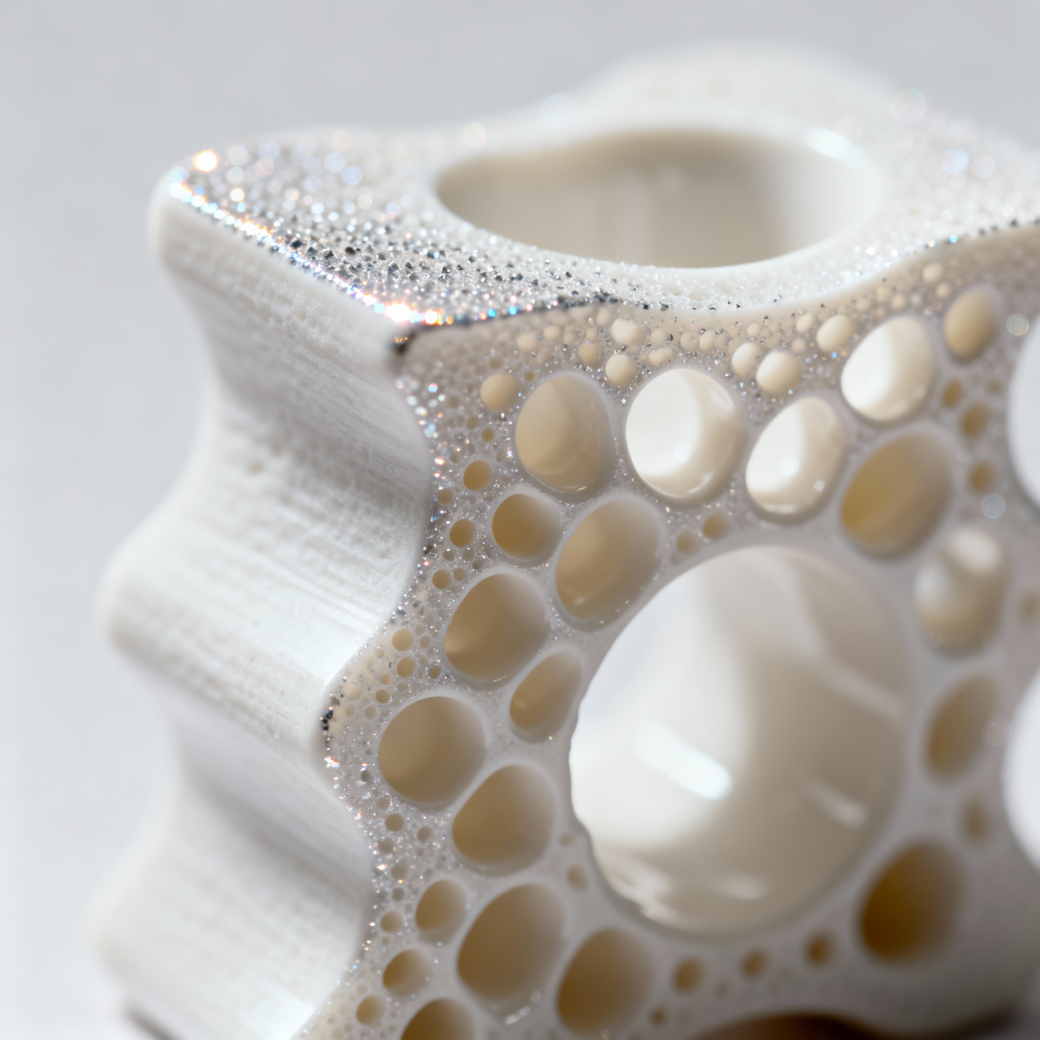
3D printing technologies for custom orthopedic implants: from design to biocompatibility
Additive manufacturing is radically transforming the production of customized implantable devices in the medical sector. The ability to design custom components starting from the patient's anatomical images makes it possible to obtain more precise solutions than standard implants, improving stability and biological integration. In orthopedic and spinal surgery, where even minimal anatomical differences can affect clinical outcomes, 3D printing is contributing to the development of highly specific implants that reduce operating times and improve functional results.
Advanced materials for 3D printing of orthopedic implants
The landscape of materials for 3D-printed orthopedic implants is expanding beyond traditional metal alloys. While titanium remains the most widely used material due to its mechanical strength and biocompatibility, interest is growing in “metal-free” implants made from non-metallic materials, capable of offering biological compatibility and comparable structural performance.
The company Nivalon has developed a customized spinal implant free of metal components, based on advanced ceramic materials and high-performance composites. This approach offers significant advantages: high compressive strength, excellent chemical stability, better radiological compatibility, and reduced interference with diagnostic systems such as MRI and CT. Traditional metal implants can in fact present issues related to interference with diagnostic imaging, the risk of metal particle release over the long term, and problems related to excessive stiffness compared with natural bone.
A key element is the use of the 3D printing technology developed by XJet, known for a process called NanoParticle Jetting, which enables the additive production of ceramic components through the deposition of nanoparticles in suspension. This technology makes it possible to obtain high-quality surfaces, complex geometries impossible with traditional methods, high density of the final material, and precision suitable for medical applications.
Ceramic materials such as zirconia or alumina are already used for dental and joint prostheses thanks to their high hardness, excellent wear resistance, biological stability, and absence of corrosion. Applying these materials also to spinal implants requires highly controlled production processes, which additive manufacturing can make more flexible and adaptable.
3D scanning and modeling processes for personalized anatomy
The process of customizing orthopedic implants begins with the acquisition of high-resolution digital images. In Vietnam, a medical team completed a complex elbow reconstruction procedure using 3D printing technologies, thanks to collaboration with the 3D printing center of VinUniversity. The process began with image acquisition via computed tomography (CT) of the patient's limb.
The data obtained were processed by technicians to create a virtual three-dimensional model of the deformed elbow. Through advanced modeling software, doctors were able to virtually simulate the osteotomy, that is, the bone cut necessary to realign the joint. This “digital twin” made it possible to identify the exact intervention points and to predict the aesthetic and functional result before even incising the patient's skin.
In Belgium, the AZORG hospital group chose to structurally integrate 3D printing through the establishment of PrintPlace on the Moorselbaan campus (Aalst). The new headquarters on the campus includes 3D visualization tools, 3D scanners, and about thirty 3D printers, with the aim of reducing external steps and cycle times for solutions that arise from concrete clinical needs. The approach enhances the “upstream” phase (co-design, engineering, testing, iterations), making it more fluid thanks to the physical proximity between clinicians and the technical team.
A practical example is the use of printed models to help communication with the patient: in some cases, patients who have undergone surgery receive a 3D replica of the affected part, for informational purposes and to support the care pathway.
Biomechanical validation and fatigue testing of implants
The biomechanical validation of 3D-printed orthopedic implants represents a crucial phase to ensure safety and clinical efficacy. In the United States, the FDA has published technical guidelines for devices made with additive manufacturing and a specific discussion paper on point-of-care 3D printing. In Europe, the general framework remains that of Regulation (EU) 2017/745 (MDR) for medical devices, while standards such as ISO 13485 are often cited as a reference for quality systems in the sector.
When a hospital produces objects related to the clinical pathway in-house, topics such as traceability, material management, process control, post-processing, and acceptance criteria become central. The ability to produce pre-operative models and customized instruments in-situ reduces dependence on foreign imports and lowers costs for patients.
A customized implant can offer better load distribution, greater immediate stability after surgery, reduced risk of misalignment, and the possibility of integrating controlled porosity to promote osseointegration. In particular, porous or lattice structures can promote bone growth within the implant, improving vertebral fusion in the long term.
Integration of porous surfaces and bioactive coatings
One of the main advantages of 3D printing in the orthopedic field is the possibility of designing implants that exactly follow the patient's morphology and integrate functional surfaces. A customized implant with porous structures can promote bone growth within the implant itself, improving biological integration in the long term.
XJet's NanoParticle Jetting technology makes it possible to produce ceramic components with complex geometries impossible to achieve with traditional methods, while maintaining high-quality surfaces and high density of the final material. This ability to produce customized ceramic structures represents a significant step forward compared with metal-only solutions.
In the case of the elbow reconstruction surgery in Vietnam, the use of biocompatible materials certified for medical use ensured the safety of contact with internal tissues. Thanks to the surgical guides produced in-house by VinUniversity, the time spent under anesthesia was reduced by 30%, minimizing the risk of infections and accelerating the post-operative recovery phase.
In recent years, several clinical cases and industrial projects have shown how 3D printing can transform spinal surgery through custom vertebral implants for tumors or deformities, optimized porous intervertebral cages, devices with better radiological compatibility, and rapid production for complex cases.
Sterilization protocols and industrial quality control
Sterilization and quality control protocols are fundamental elements when 3D printing enters the hospital setting. The use of technical ceramics in the medical field requires highly controlled production processes, which additive manufacturing can make more flexible and adaptable while maintaining rigorous standards.
Advanced ceramic materials such as zirconia and alumina offer high hardness, excellent wear resistance, biological stability, and absence of corrosion. However, these materials require specific post-processing and sterilization protocols to ensure clinical safety.
The establishment of the 3D technology center at VinUniversity represents a paradigm shift for healthcare, demonstrating how the ability to produce pre-operative models and customized instruments in situ can reduce dependence on foreign imports and lower costs for patients. In addition to orthopedics, the center is exploring applications in maxillofacial and cardiovascular surgery.
AZORG and PrintPlace describe the integration of 3D printing as a structural choice: placing skills and tools where needs arise (departments and services) to reduce friction and increase the ability to create ad hoc solutions. In a multi-site healthcare context, the outcome also depends on governance, clinical priorities, and how quality and responsibility are managed throughout the entire life cycle of printed products.
Future prospects and standardization of production processes
The creation of customized orthopedic implants through 3D printing highlights a clear direction: the surgery of the future will be increasingly oriented toward custom-designed devices made with advanced materials. The integration of medical imaging, digital design, and additive manufacturing makes it possible to develop implants that are more compatible, more precise, and potentially more effective in the long term.
The sector is exploring solutions that are not only more customized but also based on alternative materials capable of overcoming some historical limitations of metal alloys. The “metal-free” approach opens up interesting prospects, especially for spinal applications, where stability and anatomical precision are crucial.
The standardization of production processes represents the main challenge for enabling a broader diffusion of these technologies. The need to balance customization and repeatability, innovation and regulatory compliance, requires the development of shared protocols and increasingly precise technical guidelines.
The experience of centers such as VinUniversity and AZORG demonstrates that the integration of engineering and medicine can improve surgical precision and offer customized therapeutic solutions even in extremely complex clinical contexts. This type of approach could become an important reference for the next generation of orthopedic devices, with both clinical and technological benefits, paving the way for increasingly personalized medicine based on digital manufacturing.
article written with the help of artificial intelligence systems
Q&A
How does 3D printing improve the precision and outcome of orthopedic procedures?
Starting from the patient’s anatomical images, custom implants are designed that perfectly match individual morphology, reducing operating time and improving stability and biological integration compared to standard implants.
What advantages do ceramic materials offer over metal alloys in 3D-printed spinal implants?
Ceramics (zirconia, alumina) produce no MRI/CT artifacts, release no metal ions, have stiffness similar to bone and excellent biocompatibility; they also allow controlled porosity to promote osseointegration without corrosion issues.
How is anatomical customization of an implant achieved?
High-resolution CT images are acquired, a virtual 3D model (“digital twin”) is reconstructed and the surgery is simulated; the file is then sent to the 3D printer to produce the implant or surgical guides exactly calibrated to the patient.
What are the main standards and tests required to validate a 3D-printed orthopedic implant?
In Europe the MDR 2017/745 and ISO 13485 apply; fatigue tests, load distribution, osseointegration, material traceability and documented sterilization protocols are required to obtain clinical approval.
How does hospital integration of 3D printing reduce costs and treatment times?
Centers like PrintPlace or VinUniversity produce models, guides and implants in-house, eliminating external suppliers, shortening the supply chain and lowering patient costs, while also reducing anesthesia time by 30%.