How Surgical Planning Works with 3D Anatomical Models in Medical Education
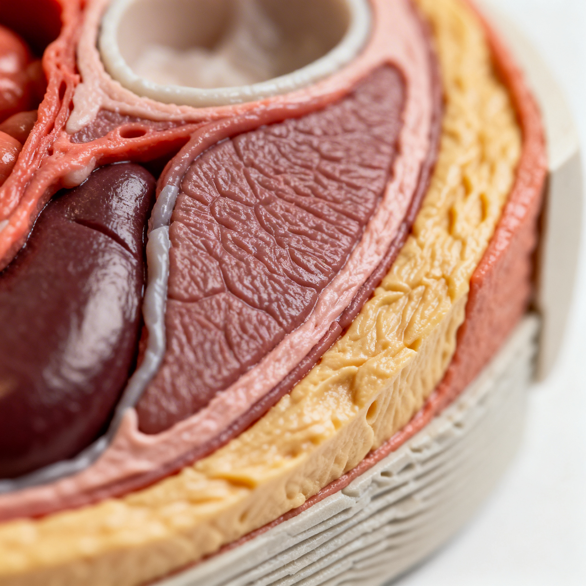
A 3D-printed facial model is revolutionizing surgeon training, enabling simulated procedures on artificial but realistically layered tissues. Thanks to the combination of advanced materials and multilayer printing technologies, 3D anatomical models allow future healthcare professionals to practice complex procedures without risk to patients, accurately reproducing the physical properties of human tissues.
The Anatomical Design of 3D Models
3D anatomical models are created by transforming radiological data into replicas faithful to clinical reality, enabling accurate and personalized surgical planning.
The process of creating anatomical models for surgical training begins with the acquisition of high-resolution images via CT or CBCT (Cone Beam Computed Tomography). These data are processed through specialized software that performs automatic segmentation of anatomical structures, completing an accurate 3D reconstruction in about an hour. AI-driven technology makes it possible to identify and separate the different anatomical components – bones, muscles, nerves, blood vessels – creating a complete digital map of the region to be replicated.
In the case of the facial models developed by Addion GmbH in collaboration with the University of Innsbruck, the anatomical design focuses on the ocular and periorbital region, replicating complex structures such as the orbicularis muscle, the orbital septum, the superior tarsal plate, the ligaments, and the facial nerve. Each element is colored distinctively in the final model, facilitating identification by surgeons in training and allowing them to understand which structures can be manipulated and which must be preserved during the procedure.
Multilayer Materials for Tissue Simulation
Multimaterial 3D printing technologies make it possible to replicate the mechanical and tactile properties of human tissues through precise combinations of specialized materials.
The most advanced anatomical models use up to eight different materials to faithfully reproduce the characteristics of human tissues. Stratasys' PolyJet technology, used in the Digital Anatomy Solution, makes it possible to combine materials with different hardness, transparency, flexibility, and resistance within the same model. Each layer is designed to respond predictably during dissection: the skin has the right resistance to cutting and maintains its connection with the underlying tissue, the muscles show the appropriate consistency, while blood vessels release a red liquid that simulates bleeding when they are incised.
The anatomical presets developed by Stratasys for dental and medical applications use predefined parameters that control how materials are mixed and distributed within the model, using specific ratios and patterns to achieve consistent and repeatable results. This standardization allows medical institutions to produce musculoskeletal and anatomical systems that biomechanically and accurately reproduce real human anatomy, while maintaining the possibility of customization based on CBCT scan data to reflect patient-specific pathologies.
Model Construction and Calibration Process
From the initial scan to the final print, the production process includes precise validation and quality control stages to ensure anatomical and functional accuracy.
Once the digital design is complete, the physical production of the model follows a rigorously controlled workflow. Polymer surgical guides can be delivered within 24–48 hours, while metal implants and complex anatomical models typically require 3–7 days for printing, post-processing, and quality assurance. All devices undergo dimensional checks and validation according to ISO 13485 workflows, ensuring that every model meets the quality standards required for educational and clinical use.
In the case of reusable models for medical education, such as the one developed at Iowa State University for meniscal injuries, the process involves 3D printing the rigid bone components and creating molds for the flexible parts. Plastisol, a flexible plastic material, is heated until it becomes fluid, poured into the 3D-printed molds, and left to solidify to produce inserts that can be mounted on the bone structures. This feature allows the material to be cut, heated, and reshaped multiple times, enabling students to practice multiple injury scenarios with the same set of components.
Educational Applications in Surgical Training Courses
3D models are used to teach complex surgical techniques, allowing students to connect anatomical theory to clinical practice safely and effectively.
By November 2025, 40 surgeons in training at the Anatomical Institute of the University of Innsbruck have used 3D-printed facial models to practice ocular and periorbital procedures. During training sessions, students perform realistic incisions, separate skin layers from the underlying muscle tissue, identify critical structures such as the facial nerve (which must not be damaged), and practice complete suturing. According to Alexander Hechenberger, CEO of Addion, “there is a specific model for almost every ocular pathology,” making medical education more accessible and practical than traditional methods based on body or animal donors.
In the dental field, Stratasys anatomical presets are used in classrooms and clinics to practice surgical techniques such as tooth extractions, implant placement, periodontal surgery, endodontic surgery, and maxillary sinus augmentation. Medical and dental device manufacturers can also use these models to accelerate clinician adoption through high-quality hands-on demonstrations and to shorten development cycles by testing instruments and techniques on anatomically accurate replicas.
Student Feedback and Assessment of Educational Effectiveness
Studies and testimonials show that 3D models significantly improve anatomical understanding and the practical preparation of future surgeons.
Student testimonials emphasize how traditional textbooks can only go so far in understanding dynamic anatomy. As reported by students at Iowa State University, “since you can’t remove someone’s leg to see inside the knee,” having a model in front of you that shows the meniscus in motion helps you understand how different stresses cause injuries and why some symptoms appear, disappear, or remain silent. Athletic Training program directors emphasize the importance of having educational tools that allow students not only to memorize definitions, but to truly demonstrate what is being discussed when describing a specific injury to a patient.
Models allow linking the “mechanism” of the injury – the dynamics of the movement that causes the damage – with the observed clinical “result” in the form of a rupture pattern, pain, joint blockage, or instability. This more intuitive mechanical understanding better prepares students to communicate with patients and make informed decisions based on solid practical knowledge, taking learning to a new level compared with traditional teaching methods.
Conclusion
3D anatomical models represent an increasingly essential tool for safe and effective medical education. The ability to faithfully replicate the physical properties of human tissues through multilayer materials, combined with the possibility of customization based on real radiological data, offers trainee surgeons an unprecedented hands-on experience. The integration of 3D imaging, digital modeling, and advanced printing makes it possible to “hold in one’s hand” complex anatomy before operating on real patients, significantly reducing risks and improving professional preparation.
Training institutions should invest in advanced simulation technologies to best prepare future healthcare professionals. With the continuous evolution of materials and 3D printing techniques, these teaching tools will become increasingly accessible and indispensable for ensuring high standards in surgical training, helping to improve the quality of healthcare and patient safety.
article written with the help of artificial intelligence systems
Q&A
What is the main advantage of 3D anatomical models in medical education?
3D anatomical models allow future surgeons to practice complex procedures safely, without risks to patients. They faithfully reproduce the physical properties of human tissues, enabling realistic and personalized training.
How are 3D anatomical models created for surgical training?
Models are obtained by transforming radiological data, such as CT or CBCT, into accurate replicas of clinical reality. Through specialized software and AI-driven technologies, automatic segmentation of anatomical structures is performed to create a precise 3D reconstruction.
What materials are used to make the models realistic?
Multi-material 3D printing technologies, such as Stratasys PolyJet, use up to eight different materials to replicate the mechanical and tactile properties of human tissues. These include materials with varying hardness, transparency, and flexibility to simulate skin, muscles, and blood vessels.
How are 3D models used in surgical training courses?
Models are used to teach complex surgical techniques, allowing students to practice incisions, suturing, and identification of critical structures such as the facial nerve. They are also employed to simulate specific pathologies and enhance practical understanding of anatomy.
What is the importance of student feedback on the use of 3D models?
Students report that 3D models provide a deeper and more practical understanding of anatomy compared to traditional textbooks. They enable visualization and manipulation of internal structures in motion, improving learning and the ability to communicate with patients.