3D-printed brain implant records neural signals and cools tissue
An international team has developed a prototype of a soft neural interface that combines electrical recording and localized cooling. The device, made using additive manufacturing techniques, was tested in animal models to explore new strategies against drug-resistant epilepsy.
A multifunctional device for neuroscience research
Researchers from the Leibniz Institute for Polymer Research in Dresden, TU Dresden and the University of Sheffield have created an experimental implant that integrates three functions into a single compact system.
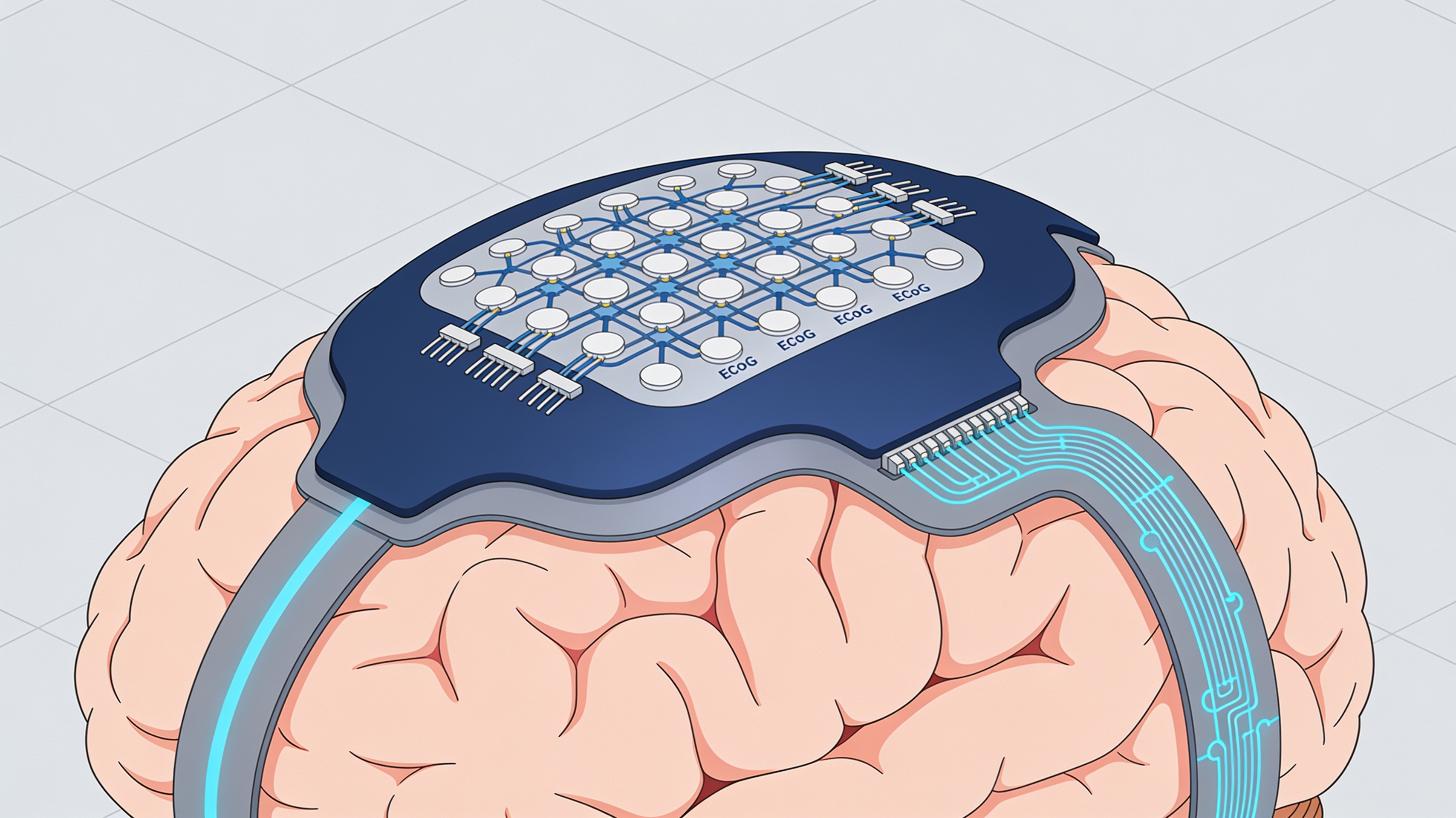
The prototype records electrical signals on the brain surface through composite electrodes. At the same time, it selectively cools an area of tissue via integrated fluidic channels. The long-term goal is to assess whether local cooling can interfere with pathological electrical activity in drug-resistant focal epilepsies.
The system is not a clinical device. It is an experimental laboratory platform, not available for medical use.
- Electrocorticographic (ECoG) recording via printed electrodes
- Local cooling via integrated fluidic channels
- Soft silicone structure compatible with brain tissue
- Tested in animal models of induced epilepsy
Additive manufacturing for complex geometries
Silicone extrusion makes it possible to organize different components in a compact volume, avoiding separate traditional manufacturing processes.
The silicone extrusion technique defines paths and interfaces in an integrated way. The process builds deformable silicone structures, fluidic channels, and supports for composite electrodes intended for ECoG recording.
Discrete electronic components complete the assembly. An external auxiliary unit controls both the thermal function and the recording function. The entire implant is not printed in a single pass.
Why cool brain tissue
The starting hypothesis is that a circumscribed decrease in temperature could interfere with pathological electrical activity without extensively cooling the brain.
The design constraint is twofold. The system must transfer heat sufficiently quickly and locally. It must also provide an electrical signal useful for verifying the effect.
Fluidic channels, polymeric materials, thermal conduction, and compatibility with the cortical surface must be designed together. The robustness of the interface represents an additional requirement.
Experimental results and limitations
The authors combined finite element simulations, physical models, and in vivo tests to validate the prototype.
Simulations and agar-based models made it possible to observe the thermal profile. In a rodent model of induced epilepsy, the printed electrodes provided ECoG recordings of quality comparable to a commercial system.
Local cooling suppressed activity resembling experimental seizures. This result does not demonstrate clinical efficacy in people nor safety of chronic implantation. The comparison concerns the conditions of the experimental protocol, not every possible disease scenario.
An uncommon element is the availability of raw in vitro and in vivo data, analysis scripts, and G-code files through TU Dresden archives. This transparency facilitates reproducibility of the results.
Prospects and obstacles to clinical translation
Moving from the experimental platform to an implantable device requires safety checks, long-term biocompatibility, and controlled clinical trials.
The availability of raw data, analysis scripts, and print files represents a transparent approach to scientific sharing. However, the gap between a working laboratory prototype and an approved medical device remains significant.
Obstacles include validation of chronic safety, miniaturization of control electronics, and demonstration of efficacy in clinical studies in patients. Additive manufacturing offers advantages in rapid prototyping and geometric customization, but does not automatically solve regulatory and clinical challenges.
article written with the help of artificial intelligence systems
Q&A
Who developed the 3D-printed neural interface?
The prototype was created by an international team involving the Leibniz Institute for Polymer Research in Dresden, TU Dresden, and the University of Sheffield.
What functions does the device integrate into a single system?
The device combines three functions: electrocorticographic (ECoG) recording via printed composite electrodes, localized tissue cooling through integrated fluidic channels, and a soft silicone structure compatible with brain tissue.
What is the goal of research on local brain cooling?
The hypothesis is that a circumscribed decrease in temperature could interfere with pathological electrical activity in drug-resistant focal epilepsies, without extensively cooling the brain.
How is the device made using additive manufacturing?
The silicone extrusion technique builds deformable silicone structures, fluidic channels, and supports for composite electrodes in an integrated manner. The implant is not printed in a single pass: discrete electronic components complete the assembly, controlled by an external auxiliary unit.
Is the device available for clinical use?
No. The system is not a clinical device, but an experimental laboratory platform tested in animal models of induced epilepsy and not available for medical use.